
Zeta Potential Measurement
Surface charge of nanoparticles — a driver of colloidal stability and cell interaction.

To determine the surface charge of nanoparticles in solution. Zeta potential influences colloidal stability, cellular interaction and transfection efficiency. Cationic (positive) charges are typical for transfection reagents to bind nucleic acids.
Zeta potential is the electrostatic potential at the slipping plane of a particle in solution. It is measured by applying an electric field and observing the velocity of particle movement (electrophoretic mobility), converted to mV.
Standard protocol
- 1
Sample preparation
- Dilute nanoparticles in filtered buffer (conductivity ~50–100 μS/cm).
- Avoid high salt, which compresses the double layer and reduces zeta potential.
- Target concentration: similar to DLS (dilute enough to avoid multiple scattering).
- 2
Cell loading
- Fill the zeta cell with ~300–400 μL of sample.
- Ensure there are no bubbles.
- 3
Measurement settings
- Temperature: 25 °C.
- Number of runs: 3–5 per sample.
- Material and dispersant settings: input the correct refractive index and viscosity.
- 4
Run measurement
- Insert the cell into the instrument.
- Apply voltage and measure electrophoretic mobility.
- Record the average zeta potential (mV).
Data interpretation
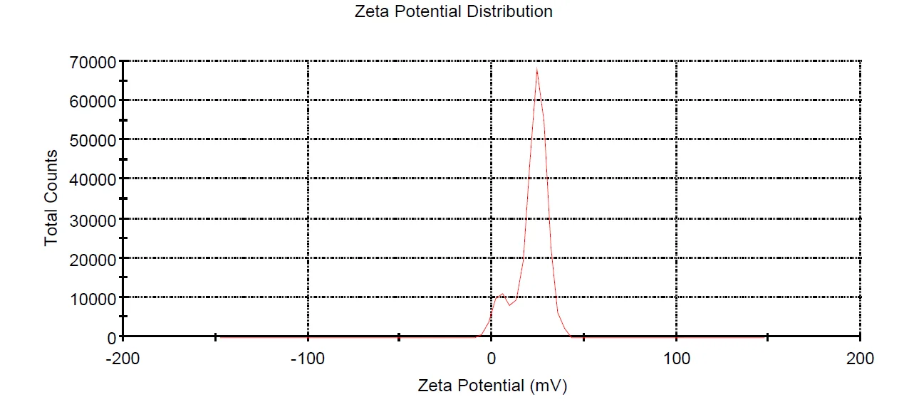
- > +30 mV or < −30 mV: high colloidal stability (strong repulsion).
- +20 to +60 mV: ideal for cationic transfection reagents (binds DNA/RNA, interacts with anionic cell membranes).
- Near 0 mV: aggregation likely.
Representative data
Let's engineer the next delivery breakthrough together
We co-develop nanocarrier and biosensing programs with pharma, biotech and academic groups — from target selection through GMP supply.