
Cytotoxicity & Biocompatibility
Annexin V – Propidium Iodide (PI) Assay
Distinguishes live, early apoptotic, late apoptotic and necrotic cells.
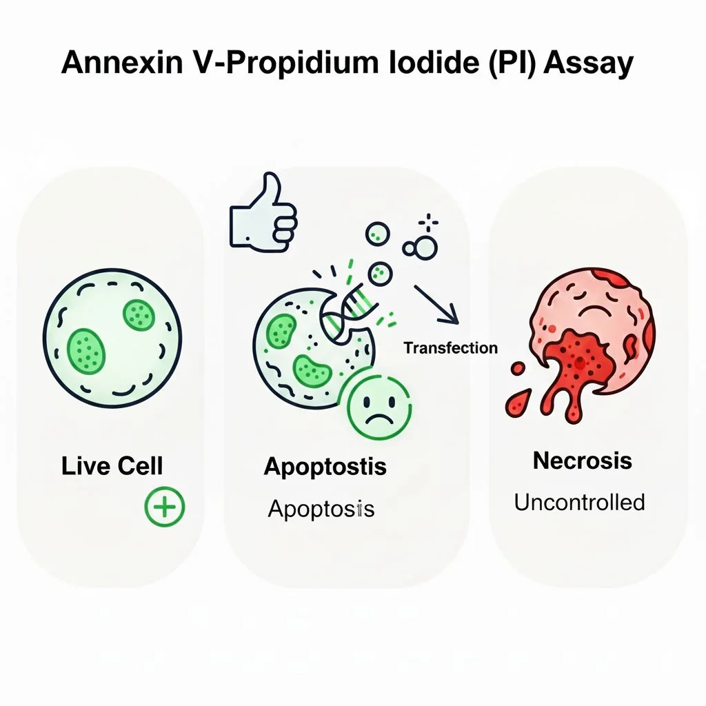
Purpose
To distinguish between live, early apoptotic, late apoptotic and necrotic cells based on phosphatidylserine (PS) externalisation and membrane integrity.
Principle
Annexin V binds to phosphatidylserine, which is exposed on the outer leaflet of the plasma membrane during early apoptosis.
Propidium iodide is a DNA stain that only enters cells with compromised membranes (late apoptotic or necrotic cells).
Standard protocol
- 1
Cell harvesting
- Adherent cells: wash with PBS, then gently detach using trypsin-EDTA (avoid over-trypsinisation).
- Neutralise trypsin with serum-containing medium.
- Collect cells by centrifugation (300 × g, 5 min) and wash once with cold PBS.
- Resuspend in binding buffer at 1 × 10⁶ cells/mL.
- 2
Staining
- Transfer 100 μL of cell suspension (1 × 10⁵ cells) to a flow tube.
- Add 5 μL Annexin V-FITC and 10 μL PI solution (20 μg/mL final).
- Mix gently and incubate in the dark at room temperature for 10–15 minutes.
- 3
Dilution
- Add 300–400 μL of binding buffer (do not wash after staining — early apoptotic cells may be lost).
- 4
Analysis
- Analyse by flow cytometry within 1 hour (do not fix).
- Controls: unstained cells, Annexin V-only and PI-only single stains.
- Set up compensation and quadrant gating.
Interpretation (quadrant analysis)
| Population | Annexin V | PI | Interpretation |
|---|---|---|---|
| Lower left | − | − | Viable (live) cells |
| Lower right | + | − | Early apoptotic |
| Upper right | + | + | Late apoptotic |
| Upper left | − | + | Necrotic (or late apoptotic with PS internalisation) |
Let's engineer the next delivery breakthrough together
We co-develop nanocarrier and biosensing programs with pharma, biotech and academic groups — from target selection through GMP supply.