
Live/Dead Assay
Two-colour fluorescence imaging of live (green) versus dead (red) cells.
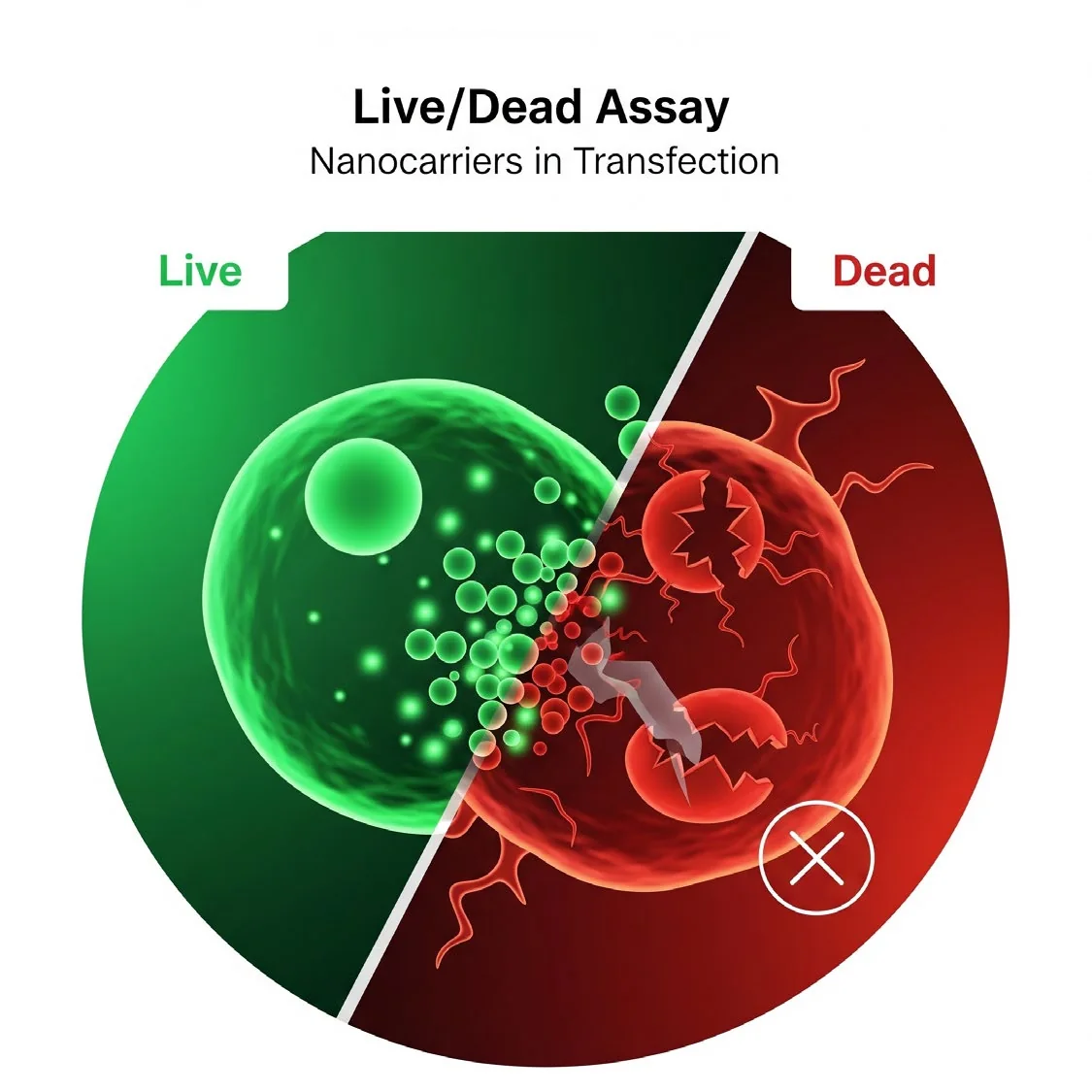
Visual assessment of viability by microscopy or flow cytometry, including 3D cultures, spheroids and tissue sections.
Calcein-AM: a cell-permeant dye converted by intracellular esterases in live cells to green fluorescent calcein (Ex/Em ~495/515 nm).
Propidium iodide (PI) or ethidium homodimer-1 (EthD-1): cell-impermeant dyes that enter dead cells with compromised membranes and bind DNA, producing red fluorescence (~535/617 nm for PI).
Standard protocol
Protocol (typical)
- 1
Wash cells with PBS or buffer.
- 2
Prepare working solution (e.g., Calcein-AM 2–4 μM + EthD-1 or PI 2–8 μM in HBSS or serum-free medium).
- 3
Add the solution to cells and incubate at 37 °C for 15–30 min.
- 4
Image using a fluorescence microscope or analyse by flow cytometry
- Green fluorescence = live cells.
- Red fluorescence = dead cells.
- 5
Avoid prolonged exposure to light.
Let's engineer the next delivery breakthrough together
We co-develop nanocarrier and biosensing programs with pharma, biotech and academic groups — from target selection through GMP supply.