
Cellular Uptake & Endocytosis
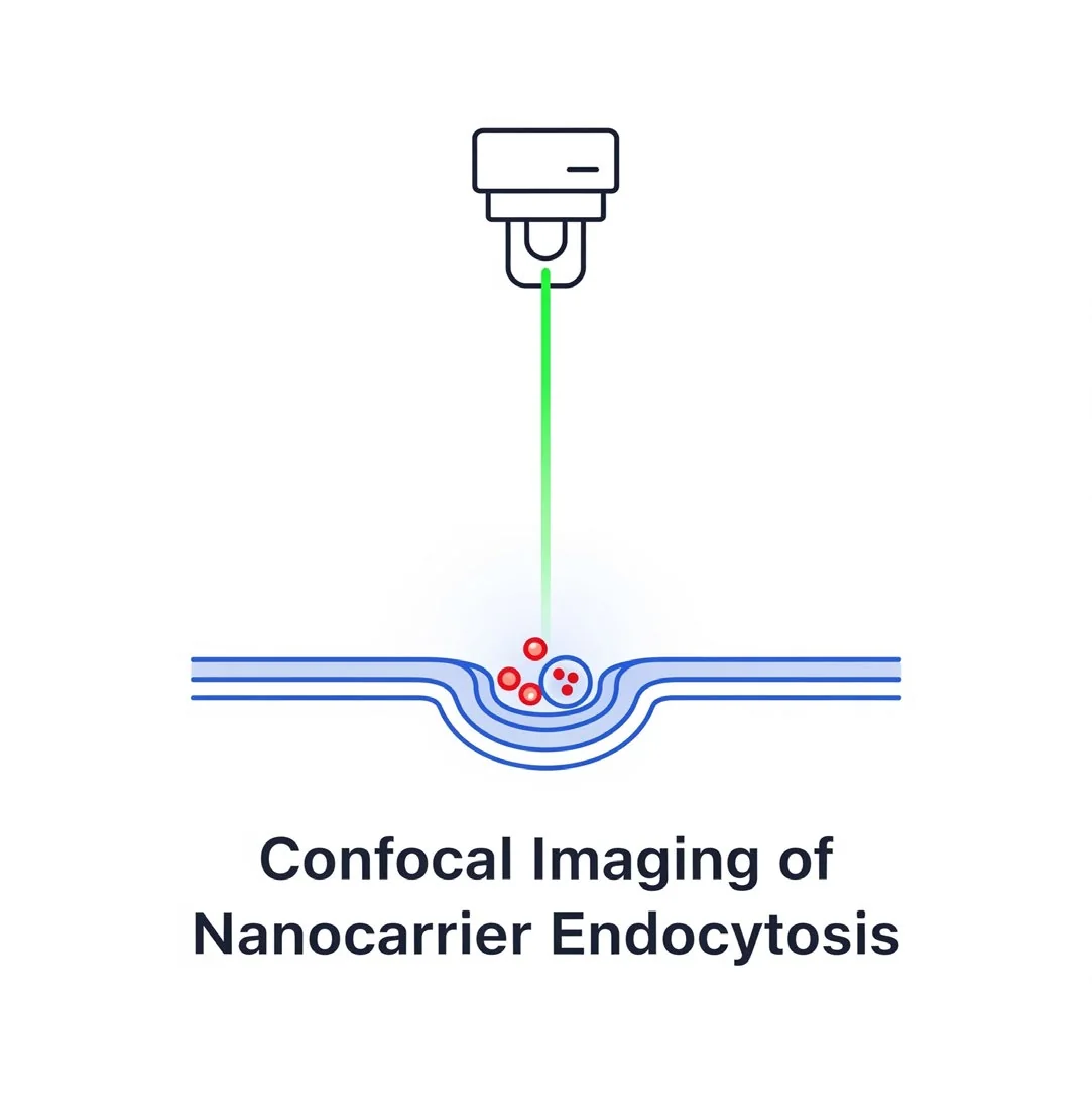
Confocal Imaging of Nanocarrier Endocytosis
Optical sectioning and 3D localisation of carriers relative to organelles.
Purpose
To obtain high-resolution, optically sectioned images of nanocarrier uptake and subcellular localisation within cells using confocal laser scanning microscopy (CLSM).
Principle
Confocal microscopy eliminates out-of-focus light, enabling 3D reconstruction and precise localisation of fluorescently labelled nanocarriers in relation to organelles (e.g., early endosomes, lysosomes).
Standard protocol
Extension of the labelled-nanocarrier protocol
- 1
Co-staining with organelle markers
- Incubate cells with LysoTracker (lysosomes), early endosome antigen 1 (EEA1) antibody, or transfect with an RFP-Rab5 plasmid.
- 2
Mounting (if needed): for fixed samples, mount with anti-fade mounting medium.
- 3
Image acquisition
- Use sequential scanning to avoid bleed-through.
- Z-stacks (0.5–1 μm steps) across cell depth.
- 3D reconstruction and co-localisation analysis (e.g., Pearson’s coefficient).
- 4
Controls
- Unlabelled nanocarriers (autofluorescence control).
- Single-stained samples (for spectral unmixing).
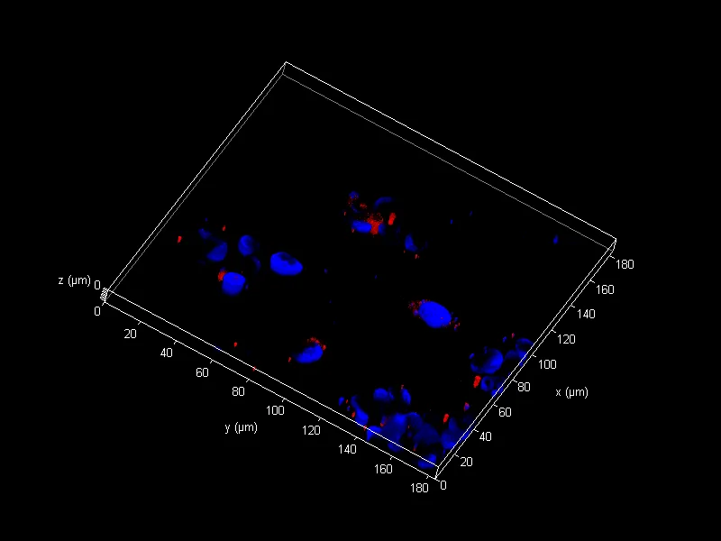
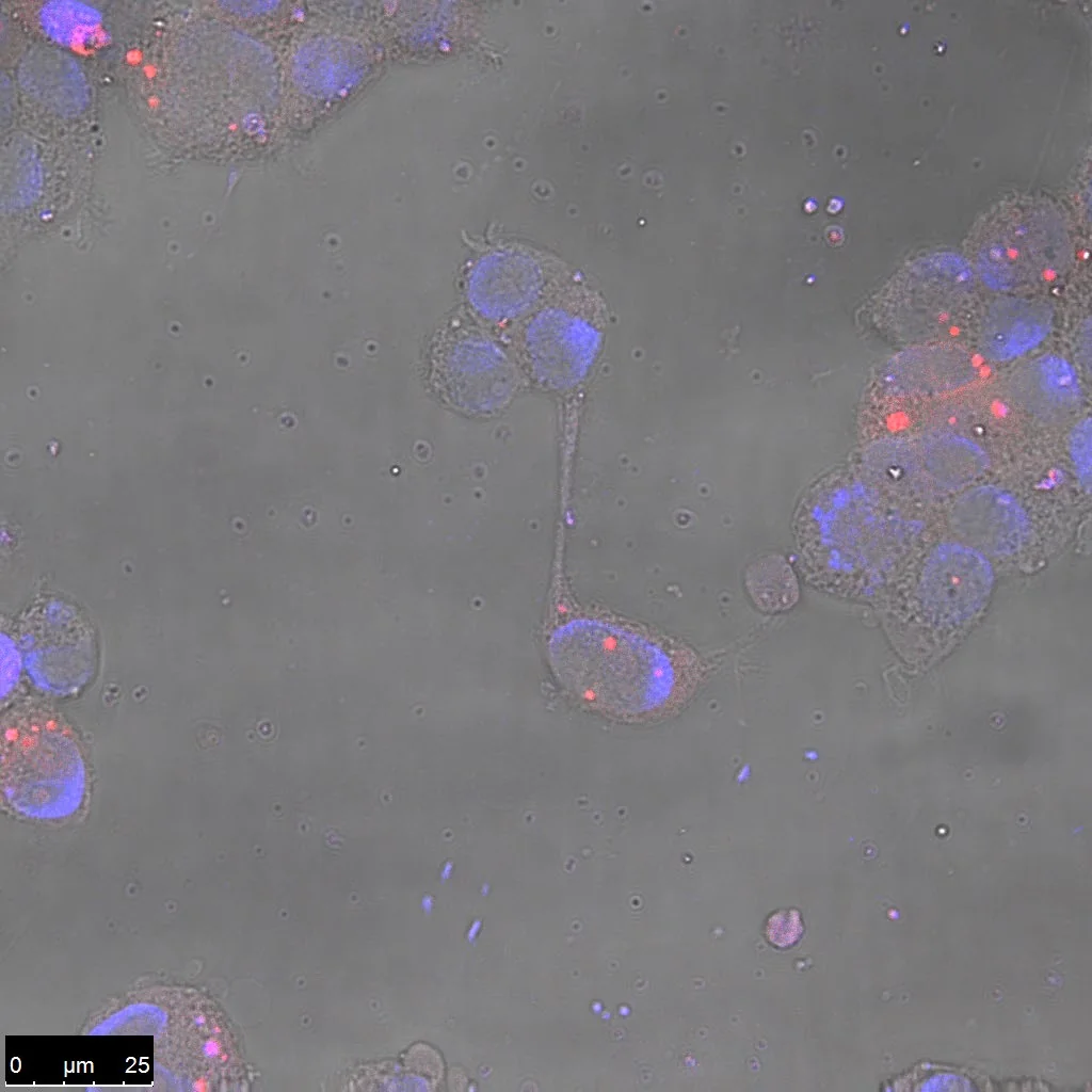
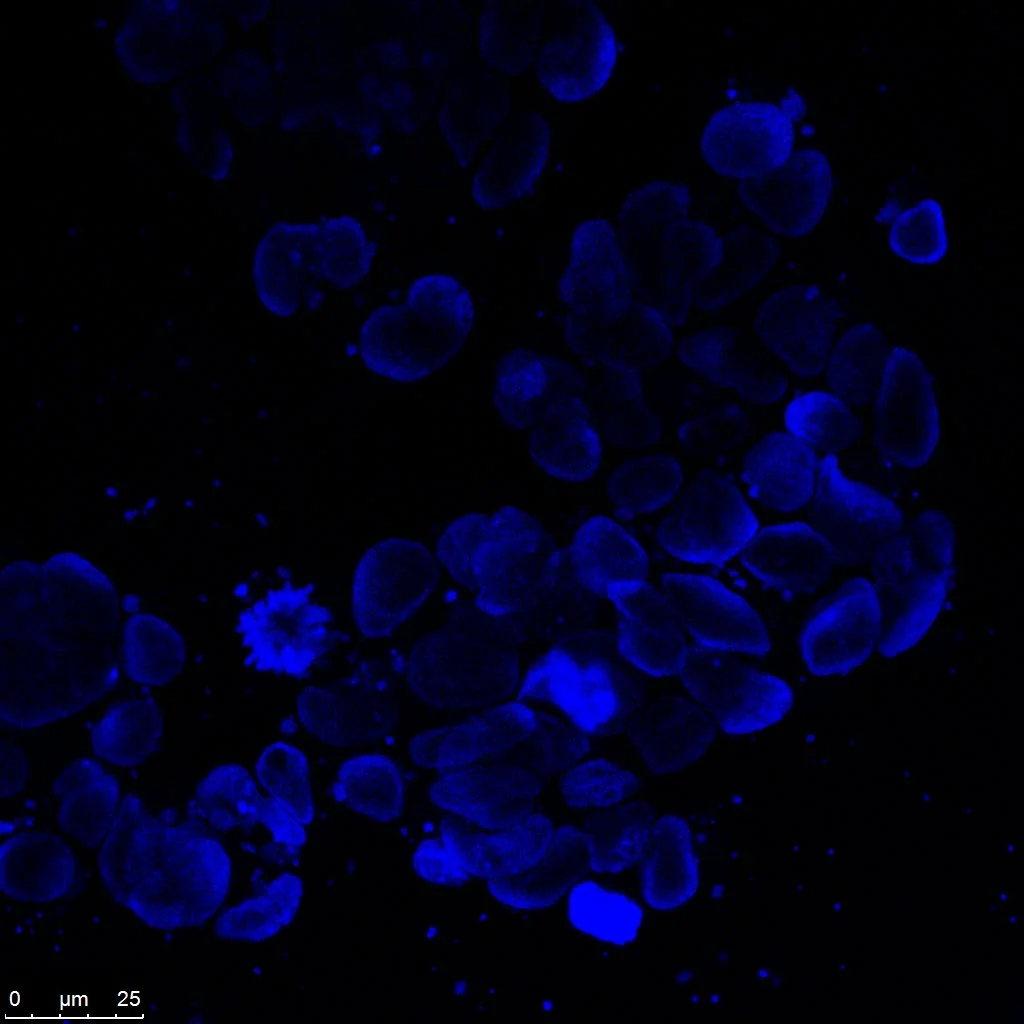
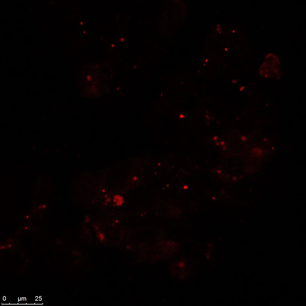
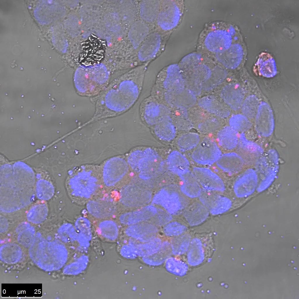
Representative data
Let's engineer the next delivery breakthrough together
We co-develop nanocarrier and biosensing programs with pharma, biotech and academic groups — from target selection through GMP supply.