
Microscopy & Microanalysis
Atomic Force Microscopy (AFM)
3D surface topography, height and roughness in air or liquid — no vacuum or stain.
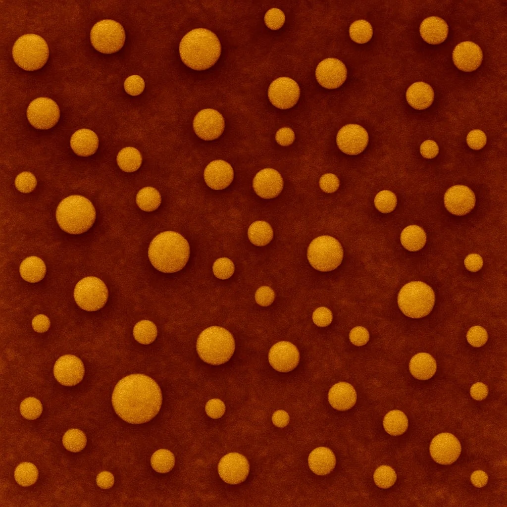
Purpose
To image surface topography in 3D at nanometre resolution under ambient or liquid conditions. Provides height, roughness and mechanical properties without requiring vacuum or staining.
Principle
A sharp probe (tip) on a cantilever scans the sample surface. Deflections due to tip–sample interactions are measured by a laser to generate a 3D topographic map.
Standard protocol
- 1
Substrate preparation
- Cut mica into ~1 cm² pieces.
- Use adhesive tape to peel a fresh, flat surface just before use.
- 2
Sample deposition
- Deposit 10–20 μL of diluted nanoparticle solution onto mica.
- Incubate 5–10 minutes for adsorption.
- 3
Rinsing
- Gently rinse with deionised water to remove unbound particles and salts.
- Tilt and drain; avoid a direct water stream.
- 4
Drying
- Air-dry or use a gentle nitrogen stream.
- 5
Imaging
- Mount the sample on the AFM stage.
- Select tapping mode (for soft samples) or contact mode.
- Scan area: 1–5 μm²; resolution: 512 × 512 pixels.
- Adjust setpoint and scan rate to minimise tip damage.
- 6
Analysis (e.g., NanoScope, Gwyddion)
- Measure particle height (more accurate than lateral size due to tip convolution).
- Generate 3D topography.
- Analyse surface roughness (RMS).
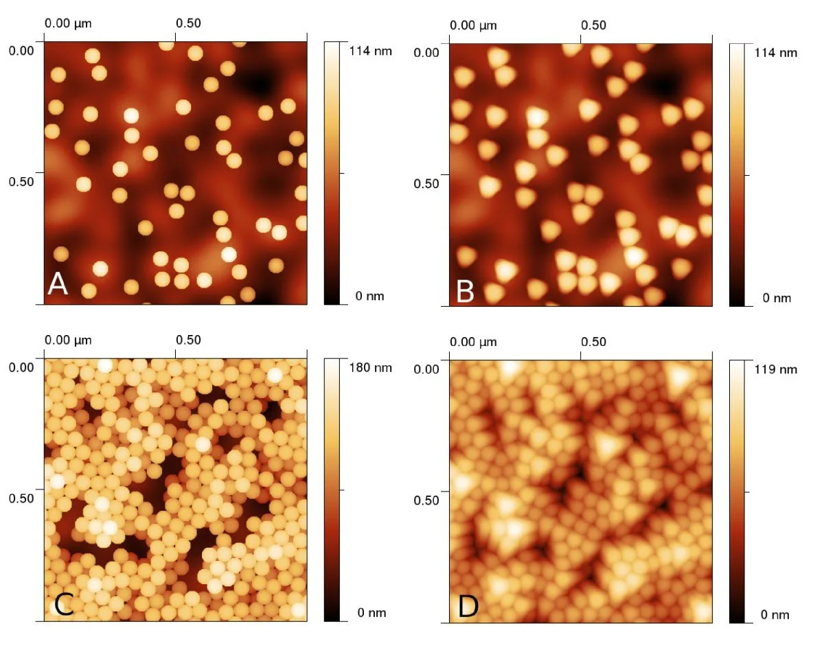
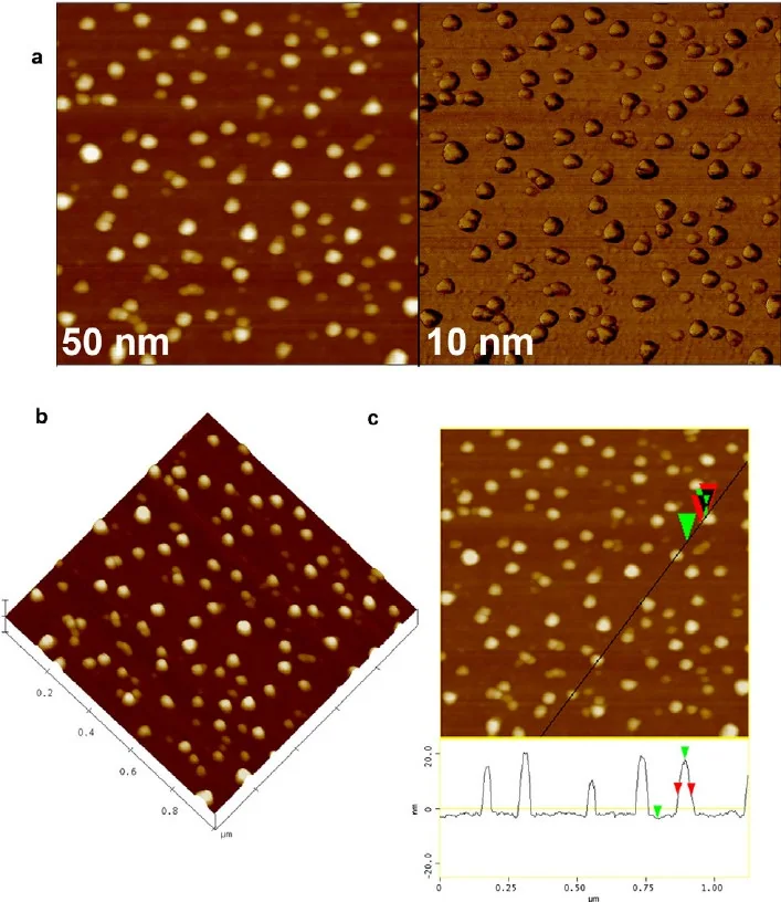
Representative data
Let's engineer the next delivery breakthrough together
We co-develop nanocarrier and biosensing programs with pharma, biotech and academic groups — from target selection through GMP supply.