
Microscopy & Microanalysis
Transmission Electron Microscopy (TEM)
Internal structure, core–shell architecture and lamellarity down to ~0.2 nm.
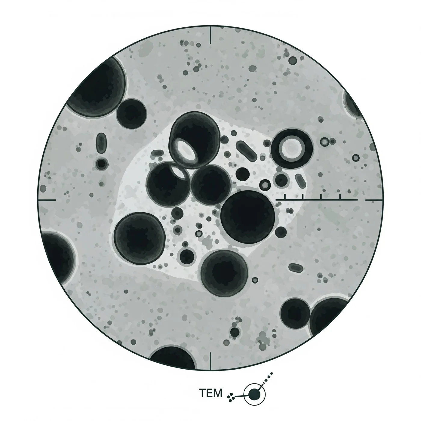
Purpose
To obtain high-resolution internal structure and morphology of nanoparticles (down to ~0.2 nm). Ideal for visualising core-shell structures, lamellarity (in liposomes) and electron density differences.
Principle
A beam of electrons passes through an ultra-thin sample. Differences in electron scattering create contrast, revealing internal structure.
Standard protocol
- 1
Grid preparation
- Glow-discharge grids (optional, improves hydrophilicity).
- 2
Sample application
- Place 5 μL of diluted nanoparticle suspension onto a carbon-coated copper grid.
- Incubate for 1–2 minutes.
- 3
Washing
- Gently blot excess liquid with filter paper.
- Wash with 5 μL of water (to remove salts), then blot again.
- 4
Staining (optional, enhances contrast)
- Apply 5 μL of negative stain (e.g., 1–2% uranyl acetate or 1% phosphotungstic acid).
- Incubate 30–60 seconds.
- Blot dry carefully.
- 5
Drying
- Air-dry in a dust-free environment or desiccator.
- 6
Imaging
- Insert the grid into the TEM.
- Use an accelerating voltage of 80–120 kV.
- Capture images at multiple magnifications (e.g., 50k×, 100k×, 200k×).
- 7
Analysis
- Evaluate shape, size, lamellarity, core-shell structure and aggregation.
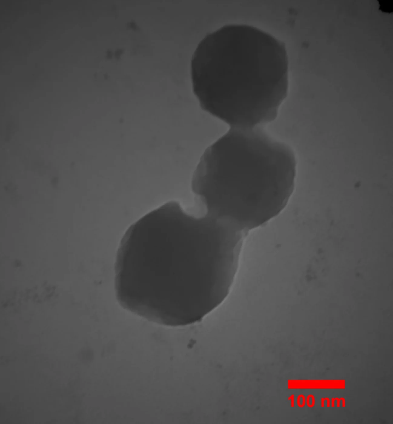
Representative data
Let's engineer the next delivery breakthrough together
We co-develop nanocarrier and biosensing programs with pharma, biotech and academic groups — from target selection through GMP supply.