
In Vitro Release Profile of Genetic Material
Release kinetics at physiological (pH 7.4) and endosomal (pH 5.5) conditions.
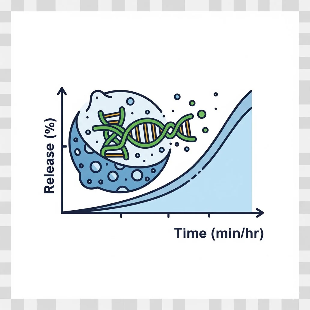
To evaluate the kinetics of nucleic acid release from the nanocarrier under simulated physiological conditions (e.g., pH 7.4) or endosomal conditions (e.g., pH 5.5), mimicking in vivo behaviour. This helps predict stability in circulation, triggered release in endosomes/lysosomes, and sustained vs. burst release.
Nanoparticles are placed in a release medium (buffer) and separated at time points using dialysis, centrifugal filtration or size-exclusion columns. The amount of released nucleic acid in the external medium is quantified (e.g., via RiboGreen® or PicoGreen®).
Standard protocol
Step 1 — Prepare dialysis system
- 1
Soak the dialysis membrane in water or buffer (as per manufacturer).
- 2
Load 1–2 mL of nanoparticle suspension into the dialysis bag/cassette and seal tightly.
Step 2 — Dialyse against release medium
- 1
Place the dialysis unit in a large volume (e.g., 20–50 mL) of pre-warmed release buffer.
- 2
Stir gently at 37 °C.
- 3
At predetermined time points (e.g., 0.5, 1, 2, 4, 8, 24, 48, 72 h), withdraw 0.5–1 mL of external medium.
- 4
Replace with fresh buffer to maintain sink conditions.
Step 3 — Quantify released nucleic acid
- 1
From each sample, mix 2 μL with 98 μL TE buffer.
- 2
Add 100 μL RiboGreen® working solution and incubate 5–10 min in the dark.
- 3
Measure fluorescence and use the standard curve to calculate released RNA/DNA.
Cumulative release (%) = (Amount released at time t ÷ Total encapsulated amount) × 100
Data interpretation
- Burst release: >30% in the first 2–4 h → rapid leakage.
- Sustained release: gradual increase over 24–72 h.
- pH-sensitive release: faster at pH 5.5 than pH 7.4 (desirable for endosomal escape).
Let's engineer the next delivery breakthrough together
We co-develop nanocarrier and biosensing programs with pharma, biotech and academic groups — from target selection through GMP supply.